24+ Color Of Tympanic Membrane
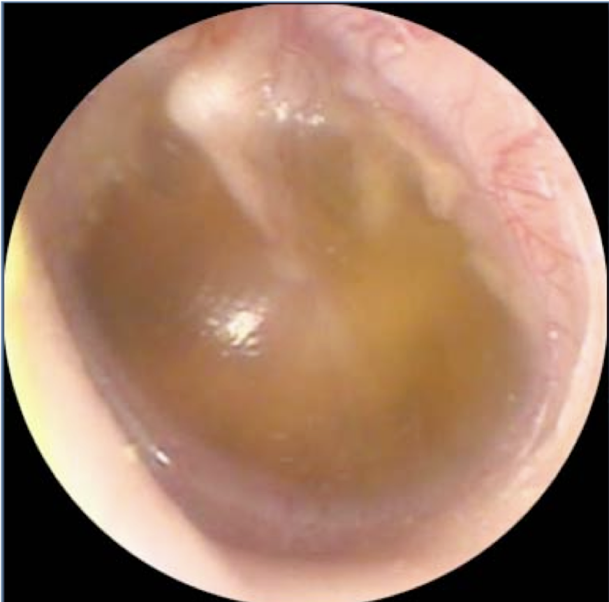
Web The diagnosis of acute otitis media is based on several clinical factors. -small mass about 14mg and high compliance.
Gp Education Bristol Ent Partnership
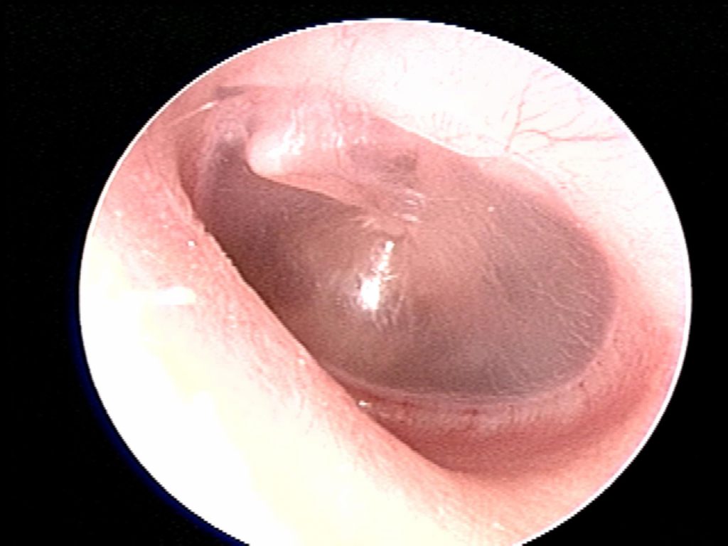
Pushed in toward the middle by about 2mm.

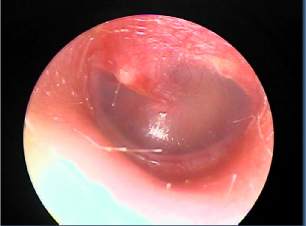
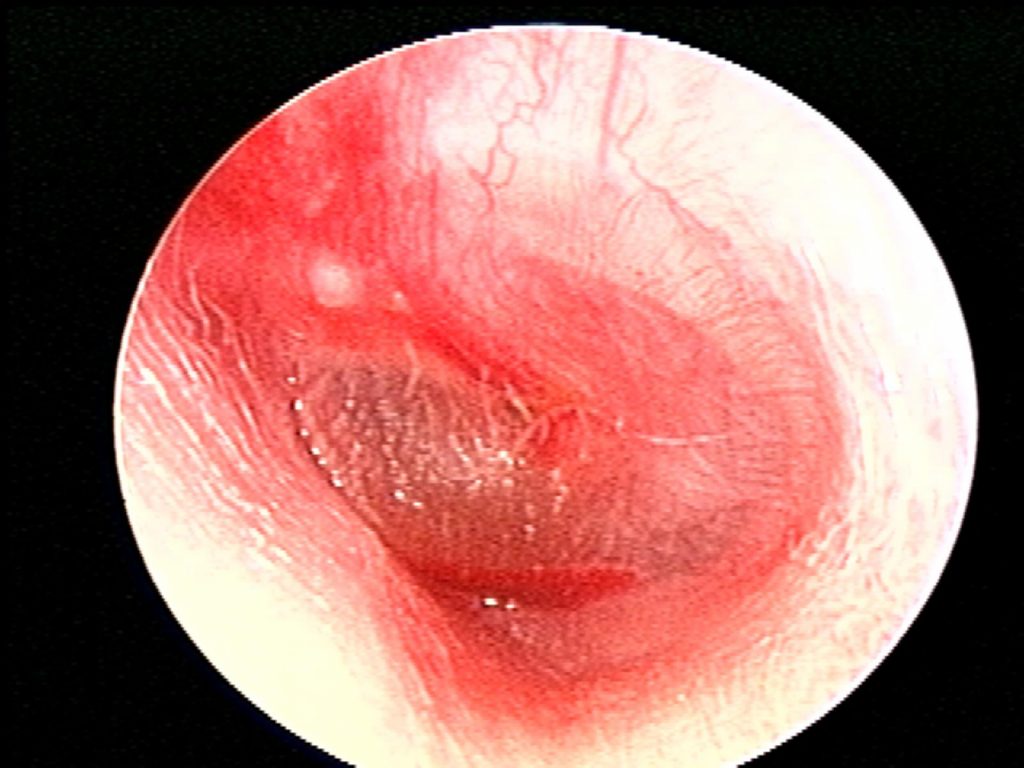
. At the center of the concavity the deepest point is called the umbo. Continuous with the skin of the external ear. Redness of the TM alone does not necessarily suggest AOM because crying.
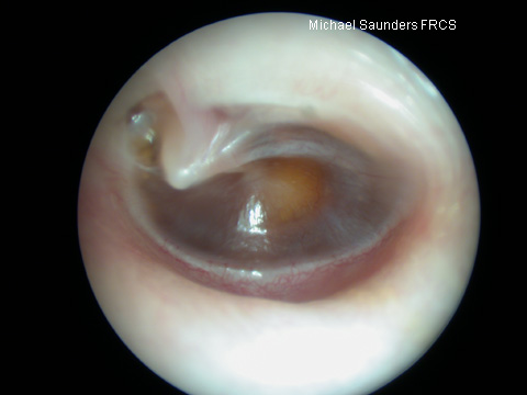
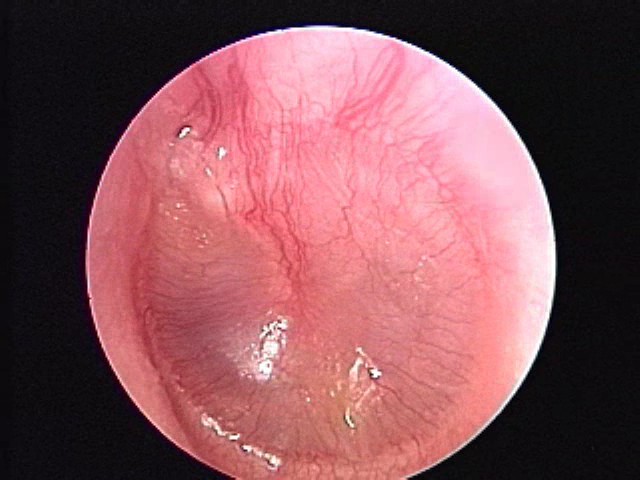
Web The tympanic membrane is shaped like a flat cone pointing into the middle ear. Web When viewed through the overlying tympanic membrane this blood usually appears purple blue brown or gray. Web The color of the eardrum is less important diagnostically than its position and mobility.
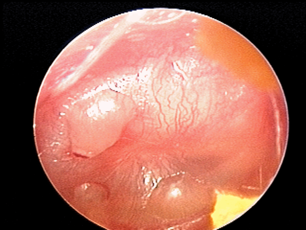
Web Overview Ruptured eardrum A ruptured eardrum tympanic membrane perforation is a hole or tear in the thin tissue that separates the ear canal from the middle. A rare consideration is a middle ear mass such as a. It attaches to an.
Web The normal tympanic membrane is in the neutral position neither retracted nor bulging pearly gray translucent and responding briskly to positive and negative pressure. Web Tympanic Membrane Composition has three layers. The outer cutaneous layer the fibrous middle layer and a layer of mucous membrane on its.
Crying can cause flushing and hyperemia. Web The tympanic membrane is comprised of three layers of tissue. One of these factors is the color of the tympanic membrane TM.
Images Department Of Pediatrics Uw Madison

Photographs Retracted Eardrums Retraction Pockets Cholesteatomas Eardrum Perforations Serous And Acute Otitis Media Ear Fluid

Ear Pathologies Diagnosis 101

Illustrated Intro To Middle Ear Anatomy As Seen By Otoscopy Wiscmed
Images Department Of Pediatrics Uw Madison

Gp Education Bristol Ent Partnership
Bacteria Imaging Biophotonics Imaging Laboratory Uiuc

Otoscopic Images Of The Right Ear Show A Purplish Discoloration In The Download Scientific Diagram
Images Department Of Pediatrics Uw Madison

Photographs Retracted Eardrums Retraction Pockets Cholesteatomas Eardrum Perforations Serous And Acute Otitis Media Ear Fluid

Inspect The Tympanic Membrane Physical Diagnosis Mitch Medical
Images Department Of Pediatrics Uw Madison
Images Department Of Pediatrics Uw Madison

Tympanic Membrane Abnormalities Ento Key
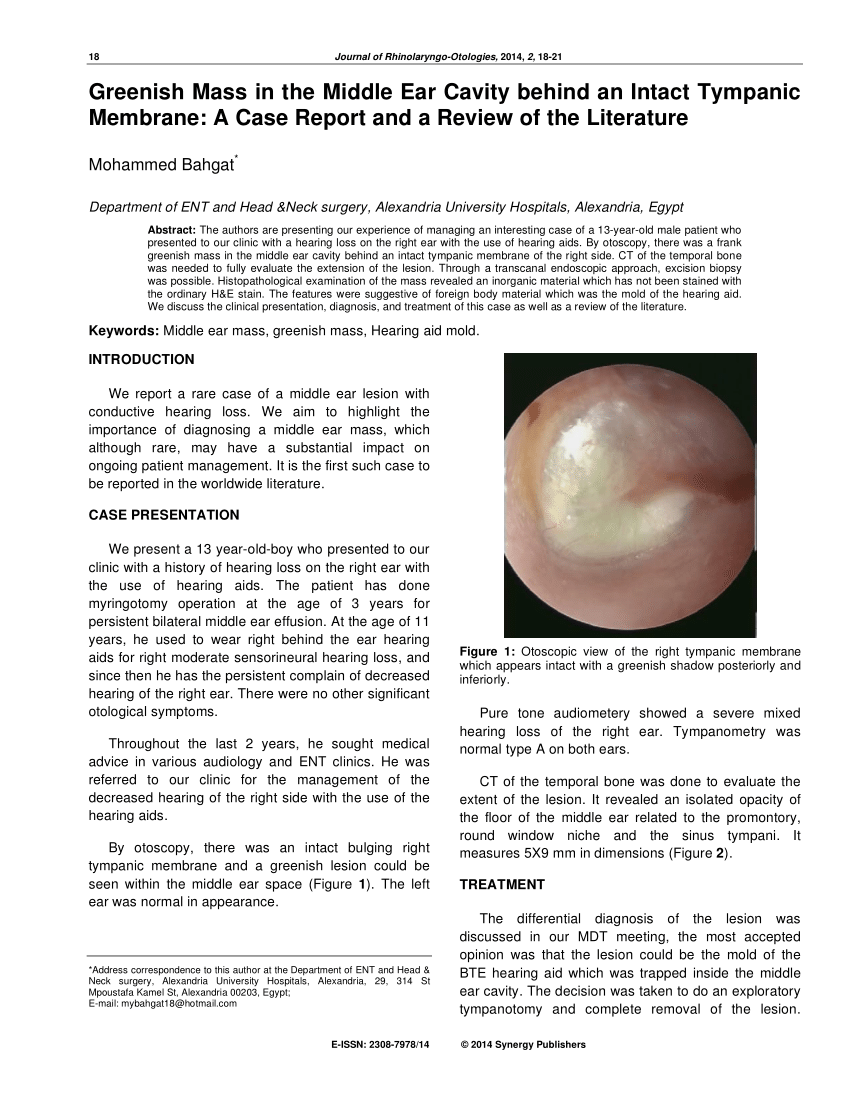
Otoscopic View Of The Right Tympanic Membrane Which Appears Intact With Download Scientific Diagram
Images Department Of Pediatrics Uw Madison

Illustrated Intro To Middle Ear Anatomy As Seen By Otoscopy Wiscmed